720-777-0123
-
Your Visit
Click to find the locations nearest you
Find locations by region
See all locations -
Research & Innovation

It starts with a Q:
For the latest cutting-edge research, innovative collaborations and remarkable discoveries in child health, read stories from across all our areas of study in Q: Advances and Answers in Pediatric Health.


Some ear deformities in children are cosmetic, while others can cause problems with your child’s hearing and development. At Children’s Hospital Colorado, our expert plastic surgeons treat a variety of ear conditions with leading-edge surgical techniques.
In addition to developmental benefits, reconstructive treatment can also help raise your child’s confidence and improve their psychological well-being. While improving your child’s hearing is always a priority, we take a holistic approach to treatment at Children’s Colorado, with the goal of not only improving physical health, but mental health as well.
What is ear deformity treatment?
Our ear deformity treatment team addresses a range of conditions that affect ear anatomy in children, including microtia, anotia and protruding ears. Whether it is using otoplasty surgery to correct protruding ears or reconstructing ears affected by microtia, our team works with each child and their family to create a custom treatment based on their unique condition and expectations.
Conditions we treat
- Microtia: A congenital (present at birth) condition that causes the external ear to be incompletely formed.
- Anotia: A congenital (present at birth) condition that causes the absence of the external ear.
- Protruding ears: Ears that stick out more than 2 cm from the head are considered to be “protruding ears.”
- Cauliflower ears: A condition that is the result of trauma, causing the ear to swell with blood or fluid.
- Earlobe deformities: Earlobes that misshaped, abnormally formed, or affected by an injury.
- Ear injuries: Traumatic injuries can range from a split earlobe to the loss of large portions of the ear, such as from a dog bite or laceration. The type of repair depends on the extent of the tissue loss.
- Stahl’s ears: A deformity consisting of an extra cartilage fold that results in a pointed ear shape.
Why choose Children’s Colorado for ear reconstruction?

Our doctors are nationally recognized experts in pediatric plastic surgery, particularly within the face, head and neck. They conducted an international study of the genetics and clinical outcomes of ear reconstruction for kids affected by microtia in Guatemala.
At Children’s Colorado, our specialists treat the whole child – not just the illness or injury. Our plastic surgery team collaborates with doctors from other pediatric specialties, including ear, nose and throat, audiology, genetics and speech pathology, to kids get the care they need. Specialists in the Microtia Clinic work together to develop holistic treatment plans for ear reconstruction for children with microtia.
Ear reconstruction treatments we offer
Otoplasty surgery for protruding ears
The surgical correction, called otoplasty, is performed by making a small incision behind the ears and by using sutures (also known as stitches) to reshape ear cartilage.
Protruding ears are characterized by ears that stick out more than 2 cm from the head. Ears that appear “pushed too far forward” are referred to as prominent ears.


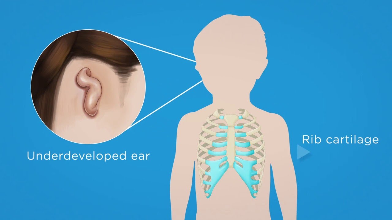
Surgical reconstruction for microtia
Microtia is a condition where the ear(s) does not fully develop while the child is in the womb. The ear is either too small or nearly absent, called lobular microtia or a constricted ear. Reconstruction for ears affected by microtia usually begins when kids are around 7 years old and involves a number of stages.
In the first stage, surgeons use cartilage from your child’s ribs to replace the missing ear cartilage. The additional stages of lobule transposition, ear elevation and reconstruction are usually completed within the following six to 12 months.