- Doctors & Departments
-
Conditions & Advice
- Overview
- Conditions and Symptoms
- Symptom Checker
- Parent Resources
- The Connection Journey
- Calm A Crying Baby
- Sports Articles
- Dosage Tables
- Baby Guide
-
Your Visit
- Overview
- Prepare for Your Visit
- Your Overnight Stay
- Send a Cheer Card
- Family and Patient Resources
- Patient Cost Estimate
- Insurance and Financial Resources
- Online Bill Pay
- Medical Records
- Policies and Procedures
- We Ask Because We Care
Click to find the locations nearest youFind locations by region
See all locations -
Community
- Overview
- Addressing the Youth Mental Health Crisis
- Calendar of Events
- Child Health Advocacy
- Community Health
- Community Partners
- Corporate Relations
- Global Health
- Patient Advocacy
- Patient Stories
- Pediatric Affiliations
- Support Children’s Colorado
- Specialty Outreach Clinics
Your Support Matters
Upcoming Events
Colorado Hospitals Substance Exposed Newborn Quality Improvement Collaborative CHoSEN Conference (Hybrid)
Monday, April 29, 2024The CHoSEN Collaborative is an effort to increase consistency in...
-
Research & Innovation
- Overview
- Pediatric Clinical Trials
- Q: Pediatric Health Advances
- Discoveries and Milestones
- Training and Internships
- Academic Affiliation
- Investigator Resources
- Funding Opportunities
- Center For Innovation
- Support Our Research
- Research Areas
It starts with a Q:
For the latest cutting-edge research, innovative collaborations and remarkable discoveries in child health, read stories from across all our areas of study in Q: Advances and Answers in Pediatric Health.


Innovative Research with 3D Technology
We collaborate with every specialty in the hospital to advance new surgical techniques, teach tomorrow’s surgeons and give our patients a brighter today.
Improving outcomes with state-of-the-art technology
The teams within the Craniofacial Center and the Cleft Lip and Palate Clinic at Children’s Hospital Colorado are using 3D technology research to advance the care of patients with craniofacial conditions. The research will also expand understanding of these conditions in the medical community.
How will 3D technology research advance care?
Previously, the results of a craniofacial surgery relied on a surgeon’s experience, expertise and a skilled eye. Now, Dr. French and team use 3D technology to study a patient’s facial features and head shape with measurements as small as a millimeter. Using the measurements, surgeons are learning how to provide better care to patients before, during and after surgery.
3D technology creates life-like 360-degree images
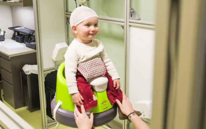
Patients pose for 3D images in a room with a specialized camera system made up of 15 cameras. The cameras capture images of the child’s face at various angles and heights. In less than one minute, the photographer takes 20 high-speed images. Then, a computer program stiches the photographs together. This creates a “life-like” 360-degree 3D image of the patient’s face and head.
Precise measurements advance understanding and outcomes
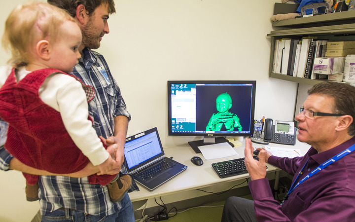
Once the 3D image is complete, Dr. French and team study the patient’s facial features like eye spacing, facial symmetry, nose length, lip shape and head shape. Surgeons then use the 3D photos to plan surgeries and as a tool for evaluating surgical results.
Benefits of 3D technology for your child’s care
The advantages of 3D technology research include:
- Precise measurements provide surgeons with an advanced level of pre-surgical planning. This planning can help achieve facial balance in early craniofacial surgeries, which can prevent additional ones.
- After surgery, surgeons see if facial balance was achieved and evaluate the improvement in head shape after a cranial vault reconstruction.
- As patients grow, surgeons will assess how a patient’s head shape and facial features change.
In addition, surgeons are developing a database of craniofacial patients at Children’s Colorado. The database will define the typical characteristics of patients with craniofacial differences (and their severity) within the hospital’s patient population.
What to expect: A painless, easy and quick technique
Participating patients and their parents enter a private room where the 3D camera system is set up. The child sits on a stool in the center of the open frame. Immediately, cameras snap photos and within five minutes, patients and their families can see the 3D image on the computer screen. Free of X-rays, the images are taken without fear of exposure to radiation. The photographs are painless and without side effects.
Conditions we evaluate
The 3D photos are used in evaluating children with conditions such as:


 720-777-0123


