720-777-0123


What if 3D rotational angiography could do the work of a CT scan?
In the Catheterization Lab at Children’s Hospital Colorado, congenital interventional cardiologists Jenny Zablah Alabi, MD, and Gareth Morgan, MD, are leveraging 3D rotational angiography, or 3DRA, in ways never described, using software and segmentation techniques Dr. Zablah largely developed herself. With them, their multidisciplinary team is creating detailed images of vessels and airways, life-size 3D printed models and virtual reality-based animations that place interventionists inside a beating heart.
New cath lab capabilities
The patient was 4 months old, weighing just 10 pounds. She was born with transposition of the great arteries. The repair was successful, but it left her with progressive severe stenosis, a common complication.
Dr. Zablah explains the procedure using a life-size 3D model she printed in her office. She points out the pulmonary arteries. They’re hardly thicker than a hairpin. A second model depicts the patient’s heart post-catheterization, detailed enough to show latticework of stents Dr. Zablah threaded up through the femoral artery and opened like tiny umbrellas at the areas of stenosis.
Dr. Morgan considers the models. “The company that manufactured our imaging system didn’t know it had this sort of capability. They didn’t even realize it was possible.”
Updated just more than a year ago with what Dr. Morgan calls “all the bells and whistles,” the system is one of the most powerful and capable on the market. Drs. Zablah and Morgan are figuring out how to leverage that capability in ways that go far beyond the commercial specs.
“We’re squeezing out every drop,” he says.
New angles of 3DRA
A staple of catheterization labs for years, 3DRA is typically used for procedural planning and sometimes guidance, layered on standard biplane angiograms to ensure accuracy.
Dr. Morgan thought 3DRA imaging could be enough. Cutting out traditional biplane imaging would mean less radiation, both for the patient and for everyone in the lab. It would mean less procedural time, a quicker recovery, less time in the hospital.
But 3DRA is tricky, a complicated setup further complicated by the fact that the heart beats. By the time contrast is injected, the team has about one second, maybe two, to get the image before the contrast floats away. Some cath teams have dealt with either by pacing the heart so fast it effectively doesn’t move blood.
Dr. Morgan wasn’t a fan of that approach. “It’s not very good for you,” he says. “Plus, if you stop the heart pumping, you’re not getting true anatomy. The heart’s pumping all the time. We want to know what it’s doing when it’s doing that.”
“It took us probably two years of trial and error,” says Dr. Zablah. “Optimizing the amount of contrast, where to give it, with which catheter, based on size and lesion.”
It helped to work in a high-volume center — the team at Children’s Colorado performs more than a thousand catheterizations every year. They tested minute variations on hundreds of cases, validating 3DRA against traditional angiograms until they knew without a doubt they were just as accurate, even better. Dr. Zablah literally wrote the protocols.
“We’re doing pulmonary valve placement with 75% less radiation than any other center,” Dr. Zablah says. “Sometimes with no contrast at all.”
Leveraging the commonality between CT and 3DRA
Their 3DRA capability got so good, so precise and so fast, in fact, Dr. Zablah started thinking about how to make it do more. Then she woke up one morning with an idea.
Technologically, 3DRA and CT have a lot in common. Both rotate X-ray around a central axis. But where CT is diagnostic and logistically cumbersome, requiring radiology support and an additional appointment of at least 45 minutes, 3DRA happens in about 5 seconds as a routine part of every cath procedure at Children’s Colorado. If Dr. Zablah could segment those images like CT scans, she reasoned, she could build detailed 3D models, even print them. They could use them to practice procedures and give them to surgeons for planning operations.
“It turned out not to be as difficult as I thought,” she says.
But she did recruit some help. She started by connecting with a computer engineer from the system’s manufacturer, who helped her write the software and override the codes necessary to make it work. She and Dr. Morgan validated the images against CT scans until they were sure their accuracy was just as good.
Then she went looking for a 3D printer.
Architectural imaging for cardiac structures
Back when Nick Jacobson was an architecture student, he gave a talk on some research he was doing in structural analysis. A surgeon happened to be in the audience. Afterward, the surgeon took Jacobson aside. If they fed CT data into the models they were using, the surgeon said, they might be able to do some fascinating things with it.
“So we gave it a try,” says Jacobson.
As it turned out, the architectural and engineering tools Jacobson was using had never been applied to medicine. They showed a lot of promise.
Today, funded by Anschutz Medical Campus innovation initiatives, his lab, Inworks, offers engineering and design support for clinician-researchers at the University of Colorado School of Medicine and at Children’s Colorado.
“We got a printer and they sent someone out to give me a one-hour tutorial,” says Dr. Zablah. “And then I started printing.”
It worked great. The models she produced were accurate and true-to-size, great for visualizing patient-specific physiology. Surgeons loved them.
3D models
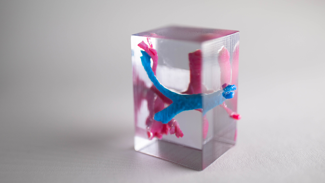
She wondered if they could print models from more pliable material her own team could use to practice placing stents. For that they used what Jacobson calls the “robust, room size printers” at Inworks. Those also came in handy when Dr. Zablah figured out how to segment not only the vessels, but the airways.
“In these babies with abnormal heart physiology, a stent can make the vessel too big and crush the airway, and then you’re in big trouble,” says Dr. Morgan. “We used to use bronchoscopes or inject contrast down the airway to figure out that anatomy. Jenny worked out a way just to pick the airway out of these pictures basically by segmenting where there’s no contrast.
“She’s the first person to work that out,” he adds.
Aside from the time and risk it eliminates, the ability to model vessels and airways simultaneously improves accuracy, since both segments come from the same data set. Working with Jacobson, Dr. Zablah color-coded those segments and started printing them in blocks of clear plastic, so they could study the intersections from any angle.
Now they’re working on virtual reality, building patient-specific data into animated models that interventionists like Drs. Zablah and Morgan can, with a set of goggles, step into and explore. That’s never been done, so it’s hard to say what insights it might yield. But the power of 3D is well established.
“A lot of studies point to the effect true 3D has on the brain,” says Jacobson. “Holding a 3D model activates a portion of the visual processing centers of the brain, responsible for problem solving, that don’t get activated any other way.”
Souvenir models for families
Back in Dr. Zablah’s office, Dr. Morgan considers a soft model of a patient’s aortic arch.
“A lot of families really don’t understand what’s wrong with their child,” he says. “But I can bring this to a family and say, this is your daughter’s aorta. It looks like this, here’s the problem, and here’s what we’re going to do. That’s powerful. When Jenny brings these models down, it’s tears, it’s hugging. It’s never been explained that way.”
Souvenir models are standard procedure for every patient now. Dr. Zablah has a drawer full of clear plastic boxes she puts them in. Sometimes she paints them. Families have used them as Christmas ornaments. Kids take them to show-and-tell.
And because Dr. Zablah wrote the software and taught herself how to do the segmentation, the entire process, from the image to the printer, takes about 10 minutes.
Precision devices for hearts of all sizes
Drs. Zablah and Morgan’s precision and expertise allows them to be a part of clinical trials for new devices that expand the capabilities of catheterization for more and more patients, avoiding high-cost, high-risk, potentially traumatizing open-heart surgeries.
One device currently being trialed is an hourglass-shaped stent interventionists can use to seat a percutaneous valve with a diameter of 29 millimeters in a distended vessel of up to 40 mm. That’s particularly useful for tetralogy of Fallot, where persistent regurgitation of blood from the pulmonary arteries can lead to severe distension of the outflow tract.
On the other end of the spectrum, they’re using a tiny device for closing the patent ductus arteriosus in the very premature. It’s a delicate operation, but Dr. Zablah successfully placed one in a baby weighing just 640 grams, the smallest in Colorado.
Featured Researchers

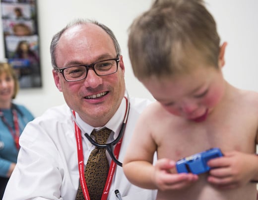
Jenny Zablah Alabi, MD
Interventional cardiologist
The Heart Institute
Children's Hospital Colorado
Associate professor
Pediatrics-Cardiology
University of Colorado School of Medicine
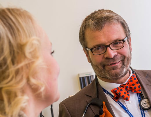
Gareth Morgan, MD
Congenital interventional cardiologist
The Heart Institute
Children’s Hospital Colorado
Professor
Pediatrics-Cardiology
University of Colorado School of Medicine