How is 3D intracardiac echocardiography (ICE) changing the standard of care for cardiac catheterizations?
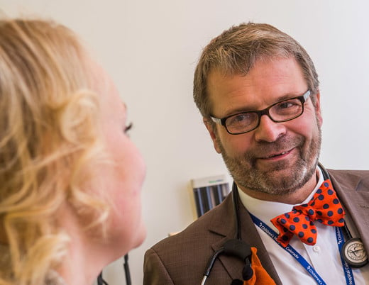
In December 2021, Children’s Hospital Colorado became the first and only pediatric hospital in the world to start using 3D ICE during cardiac catheterization procedures for pediatric patients with congenital heart disease. Led by congenital interventional cardiologist Gareth Morgan, MD, the Cardiac Catheterization Lab, or cath lab, at Children’s Colorado is at the forefront of innovation with superior cardiac visualization.
Innovation in the cath lab
Innovation is a common theme at Children’s Colorado, and that is especially true in the Heart Institute. Dr. Morgan, who recently developed a newly FDA-approved G-Armor Stent, played a major role in obtaining 3D ICE technology for the cath lab. The recently FDA-approved 3D ICE catheter from Phillips will allow cardiac interventionalists to better navigate complex cardiac procedures without any significant additional time, risk or access points.
“This was an enormous amount of effort — over 18 months — to ensure we had the pieces in place to add 3D ICE to our suite of tools,” Dr. Morgan says. “The desire of our physicians to offer the best possible tools is constant — it’s how our cath lab functions in the first place.”
As a high-volume valve center offering comprehensive imaging with existing expertise in 3D imaging, the cath lab was already positioned to obtain a 3D ICE probe for pediatric patients. And not long after the Mayo Clinic performed the first procedure (a left atrial appendage occlusion) using its 3D ICE catheter, the cath lab at Children’s Colorado introduced 3D ICE to its arsenal of high-tech imaging solutions — a testament to its reputation for driving innovation and providing some of the most comprehensive cath lab imaging.
Superior visualization with less risk
While transesophageal echocardiography (TEE) has served as the foundation for guiding structural heart interventions for most cath labs, the introduction of ICE technology represents a major advancement in cardiac imaging — and the upgrade to real-time 3D visualization takes it to another level. While most echocardiography is performed outside the heart with transthoracic echocardiography or through the esophagus with TEE, 3D ICE produces a real-time, 3D computer model of a child’s heart, offering detailed views of the heart’s complex anatomy, blood flow, interventional tools and substructures, such as percutaneous pulmonary valves that aren’t seen well with other imaging modalities.
With just a slightly larger catheter diameter, the new 3D ICE catheter uses a miniaturized ultrasound with a 3D echo probe to navigate to the center of the patient’s heart via the same vasculature used during minimally invasive cardiac procedures.
Unlike TEE, ICE can be performed without the risk of esophageal trauma and without the need for general anesthesia, which carries its own risks. Therefore, ICE can be a suitable alternative for patients who would otherwise not be good anesthesia candidates. In addition, ICE reduces fluoroscopy exposure, lowering the amount of X-ray radiation to which a patient is exposed.
With the emergence of 3D ICE, the cath lab is also reducing the number of angiograms and the iodine-based contrasts to assess valve function, decreasing damage to patients’ kidneys and thyroid glands. Despite all these minimizing steps, there is increasing evidence that ICE is the most accurate modality available in our field.
Advancing the standard of care for valve repair and replacement
While conventional ICE has largely replaced TEE for guiding certain procedures — many performed as open-heart surgery just a few years ago — it is playing an emerging role in minimally invasive valve replacement and repair. When we add 3D to a regular assessment with ICE, the result is a huge step forward.
“If we know exactly what a newly implanted valve looks like in glorious, 3D detail,” Dr. Morgan says, “then any assessment of the valve afterward can refer back to this baseline study to determine if the valve is losing function, becoming infected, narrowing or leaking.”
The higher resolution images obtained with the 3D ICE probes allow the team to put together a more thorough baseline assessment, which allows for better longitudinal follow-up, improved diagnostic accuracy and earlier recognition of complications, including valve failure, endocarditis and infections.
Future of cardiac catheterization
While 3D ICE won’t necessarily replace other imaging modalities due to its high cost, Dr. Morgan estimates that about 10% of the more than 1,000 cardiac catheterizations performed each year at the Children’s Colorado’s Heart Institute, will use 3D ICE, including all valve repairs and replacements.
As the cath lab performs more 3D ICE catheterizations, Dr. Morgan hopes the data gathered adds to the general understanding of why valves fail or deteriorate over time and where providers should focus therapy to keep valves healthy for longer.
As ICE technology improves, Dr. Morgan will continue to explore innovative ways to ensure the cath lab provides the most comprehensive imaging possible. Dr. Morgan adds, “Each day, we are exploring new ways to provide the most appropriate and effective care with the best imaging technology we can get our hands on.”
Featured Researchers

Gareth Morgan, MD
Congenital interventional cardiologist
The Heart Institute
Children’s Hospital Colorado
Professor
Pediatrics-Cardiology
University of Colorado School of Medicine


 720-777-0123


