720-777-0123


Key takeaways
-
Our researchers sought to distinguish tissue-destroying eosinophils from tissue-dwelling eosinophils that maintain homeostasis and protect against disease.
-
They identified subgroups of eosinophils based on surface marker expression.
-
The findings provide new insights into eosinophil localization, plasticity and function in acute and chronic disease.
-
Eosinophilic gastrointestinal diseases (EGIDs) are becoming more common
-
Eosinophilic esophagitis (EoE) is most common EGID
Background: Subgroups of eosinophils contribute to homeostasis or disease
Eosinophils are white blood cells produced in bone marrow. They spend a short time in the bloodstream and then move into tissues, including the gastrointestinal (GI) tract, lungs, adipose tissue, and thymus.
Eosinophils make up less than 5 percent of white blood cells in healthy people. In the GI tract, eosinophils are found in greater numbers, where they:
- Contribute to innate and adaptive immunity
- Remodel and repair tissue
- Maintain metabolic homeostasis
- Defend against pathogens

Some subgroups of eosinophils contribute to homeostasis, while others cause disease. This distinction can be leveraged to treat disease as new eosinophil-targeting biologics are developed.
Mice studies have shown two eosinophil subgroups in the lungs:
- SiglecFint
- SiglecFhi
GI eosinophils are a highly heterogenous population. CD11c expression cannot be used alone to differentiate “helpful” from “harmful” eosinophils because the marker is also found on intestinal eosinophils in the steady state.
In this study, researchers from the Gastrointestinal Eosinophilic Diseases Program in the Digestive Health Institute at Children’s Hospital Colorado sought to characterize eosinophils by subgroup, using the expression of CD11c as a framework.
Methods: eosinophils from allergen-exposed and control samples collected, prepared for subgroup identification
Mice were sensitized to ovalbumin (OVA), a food allergen implicated in egg-induced anaphylaxis and atopic dermatitis, admixed with alum adjuvant. Sensitized mice were challenged by oral administration of OVA.
OVA was replaced with phosphate-buffered saline in control mice. In some experiments mice were also pulsed with BrdU to label eosinophils generated within a small timing window, to enable tracing of the same population of eosinophils over time.
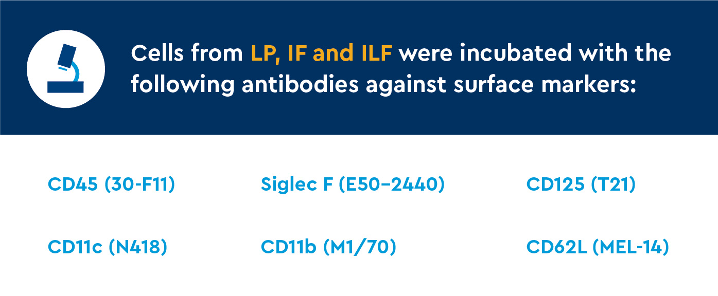
- Small intestine samples from five to six jejunal rings were fixed and stained for microscopic and immunofluorescent (IF) analysis.
- Single-cell samples from the lamina propria (LP) and intraepithelial (IE) compartments of small intestine were purified, and leukocytes were collected.
- Leukocytes were collected from intestinal lavage fluid (ILF) from the intestinal lumen.
Results: Eosinophil subgroups show plasticity after allergen exposure
Surface marker expression in eosinophil subgroups
CD11c expression overlaps were found in IE and LP cells. Gates were assigned to divide eosinophils into subsets:
- CD11c-/lo is higher in LP compartments (31% LP vs 3% IE)
- CD11cint is found in both compartments (52% LP vs 30% IE)
- CD11chi is higher in IE compartments (14% LP vs 66% IE)
Siglec F and CD11b expression in LP and IE cells increased from CD11c-/lo to CD11cint to CD11chi subpopulations.
- Compared to LP cells, IE eosinophils expressed higher levels of:
- Siglec F
- CD11b
- CD11c
Interleukin-5Rα (IL-5Rα) was uniformly detected across all CD11c-defined subgroups, and CD62L was not found uniformly in all subgroups.
Tracking CD11c-defined subsets in allergen exposed and control mice
A novel subset of the newly recruited eosinophils failed to upregulate CD11c:
- Upregulated other markers of activation (i.e., CD11b)
- Transiently observed in the IE layer on day 4
- Transient CD11c-/lo, CD11bhi population underwent transepithelial migration and was recovered in the lumen on day 4
Discussion: Eosinophils upregulate CD11c and migrate into villi in response to inflammation
Study authors discussed their approach and key findings, including:
Developing a novel CD11c-defined gating framework
Relative CD11c expression was used as a novel framework to stratify eosinophils into subsets and tracked it along with other markers of eosinophil activation:
- Intestinal eosinophils predominantly found in the LP compartment when healthy
- Small subset found in the IE compartment
- CD11c expression changed along a continuum from LP compartment to IE compartment
- Highest CD11c expression in IE compartment
CD11c-/lo represented a less activated state than CD11cint < CD11chi subpopulations based on:
- Lower Siglec F (cell surface protein that binds sialic acid)
- Lower CD11b (an adhesion molecule upregulated in asthma)
- Higher SSC (indicator of normal density; characteristic of non-activated eosinophils)
In addition, LP compartment was enriched for CD11c-/lo, IE compartment was enriched for CD11chi
All three subgroups of eosinophils are likely vulnerable to anti-IL-5 biologics, at least at baseline
- IL-5Rα was uniformly found in all subgroups
Unlike baseline lung eosinophils, intestinal eosinophils from all CD11c-defined subpopulations were uniformly negative for CD62L
Tracking CD11c-defined subsets in situ in allergen-exposed and control mice
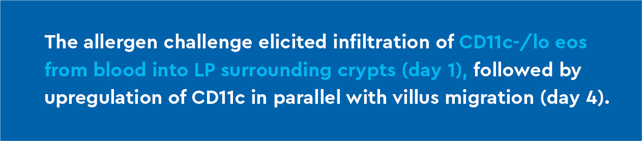
Oral allergen challenge elicited infiltration of CD11c-/lo eosinophils followed by their redistribution into villi and upregulation of CD11c:
- Increased BrdU+ CD11c-/lo eosinophils found in LP surrounding crypts (day 1), corresponding to migration from the bloodstream.
- Absolute eosinophil counts remained stable between day 1 and day 4
- BrdU+ CD11c-/lo eosinophils migrated to intestinal villi and exhibited increased CD11c expression (day 4)
- Suggests eosinophils in LP surrounding crypts upregulated CD11c expression while redistributing to intestinal villi in response to ingested allergens
- BrdU+ studies suggest these are newly recruited BrdU+ CD11c
- BrdU+ CD11cint and CD11chi eosinophils still detected within LP and IE compartments on day 11, significance unknown
In human and animal models of allergic airway inflammation, eosinophils migrate from the lung parenchyma to the airways. Researchers tested to see if eosinophils similarly migrated across the intestinal epithelium:
- CD11c-/lo eosinophils found in the ILF after allergen exposure (day 4) more closely resembled eosinophils from LP than IE compartments
- Timing and phenotype of ILF eosinophils (day 4) suggest they represent the transient population of CD11c-/lo eosinophils transiently detected in the IE compartment on day 1.
- Oral OVA challenge drove eosinophil recruitment, villus redistribution and CD11c activation
To determine whether chronic inflammation would affect the balance of CD11c-defined eosinophil sub-phenotypes, researchers used mice with a genetic deletion causing tumor necrosis factor-α (TNFα) overexpression to model chronic inflammation:
- Mice with TNFα overexpression had chronic intestinal inflammation and ileitis with increased LP CD11cint and CD11chi eosinophil subgroups
- Suggests inflammation drives changes in CD11c expression
Conclusion: Eosinophil subgroups show differences in localization, plasticity and function
This study provides a framework to track eosinophil subgroups in health and disease and provides insights into eosinophil localization, plasticity and function in acute and chronic diseases. These findings can lead to better EGID identification, tracking and treatment options as more eosinophil-targeted biologics become available.