720-777-0123


What can the medical community learn from a rare and risky surgery to resect a fetal pericardial teratoma in utero?
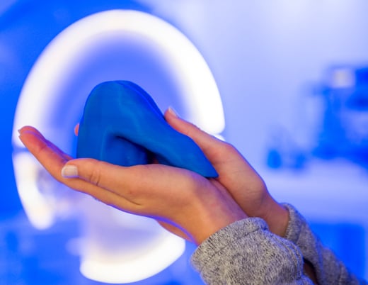
In January 2022, providers from across Children’s Hospital Colorado gathered in an operating room in the Colorado Fetal Care Center to save not one life, but two. A mother from Omaha, Nebraska had made her way to Colorado after learning that her 27-week-old fetus had a large mass pressing against its heart and needed immediate care. The mass threatened to send the fetus into hydrops, or heart failure, and carried additional risks to the mother’s health. The team carefully weighed their options and decided the best course of action was to complete the resection while the fetus remained in utero.
Facing a difficult decision
Even before the patient sought initial care at her local hospital, the groundwork for this unique surgery was already laid thanks to the relationships Bettina Cuneo, MD, a fetal cardiac specialist, had formed with providers there. So, when this difficult case arose, the patient’s doctors referred her to the team at Children’s Colorado, knowing they’d be able to provide the best care for this rare defect.
“Part of the rarity of this case was the combination of the disease and how it affects the baby,” says James Jaggers, MD, a cardiac surgeon at Children’s Colorado who helped plan and complete the resection. “It probably happens more than we know, but this fetus and this family were lucky enough to be plugged into a situation where they could get to [Dr. Cuneo] and then get to us for intervention.”
When the patient arrived in Colorado, doctors faced a difficult choice due to the young gestational age of the fetus: “Do we deliver early, do the operation and leave the baby out, or do we do the operation with the fetus still attached to the mother and put the baby back in to continue growing?” Dr. Jaggers recalls.
A rare and risky option: in utero resection
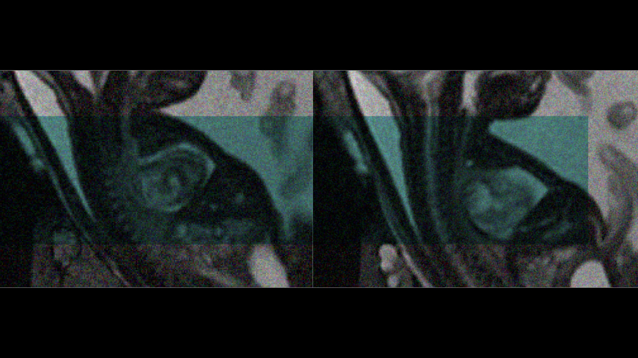
Based on the fetus’s significant prematurity and the tumor’s large size, the team decided to pursue an option that’s only been documented a handful of times in the literature: in utero resection. That, according to Drs. Cuneo and Jaggers, would give the fetus the best chance of survival. After all, there’s no better ICU for a fetus than a mother’s uterus, says Kenneth Liechty, MD, a fetal surgeon who played a critical role as a co-surgeon in this case.
Though the resection itself is a relatively common surgery, this unusual element, in which the fetus would be partially removed from the uterus for the operation and then placed back inside, generated a new level of complexity. Instead of simply removing the tumor from a delivered baby, the team would have to carefully monitor both the health of the mother and the fetus and draw a delicate balance between each patient’s needs.
“This is one of the situations where there’s a potential 200% mortality,” Dr. Jaggers explains. “You’re not just operating on one patient — you’re operating on two. And so, you always have to make sure that the safety of both of those patients is paramount.”
With that plan in mind, Drs. Jaggers, Cuneo and Liechty assembled a large team spanning multiple disciplines, from neonatology, maternal fetal medicine and cardiology to anesthesiology and obstetrics. It was vital that the team included experts who could be on hand for any complications that might arise. And of course, even more team members worked behind the scenes to make this surgery possible, including social workers and more.
“This is sort of the Super Bowl of multidisciplinary care, if you will,” Dr. Jaggers says. “It involves a lot of different services and a lot of highly specialized physicians, nurses and anesthesiologists.”
Removing the tumor during partial delivery
The surgery began with an obstetrician delivering the fetus partially outside the uterus, exposing only the arms and chest. This gave the team intravenous access to the fetus to administer anesthesia and ensure the heart would remain properly loaded with a constant supply of blood during the surgery. It also allowed them access to the tumor, which they were able to remove while the fetus remained connected to the placenta and uterus. With the tumor removed, the team was happy to find that the fetus’ heart was functioning normally.
Both patients remained stable and in good health throughout the surgery, allowing the team to complete their tasks without incident. Once they addressed the tumor, physicians carefully repositioned the fetus back in the uterus, added fluid to help avoid preterm labor and closed the uterus so both patients could begin the recovery process.
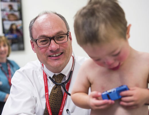
For nearly two weeks after the surgery, the fetus remained in utero before being delivered. Removing the tumor made such an impact on the baby boy’s heart that he only required a few days of care in the cardiac ICU before being transferred to the NICU to finish growing and developing.
In addition to saving two lives, the surgery has the potential to have an impact beyond this one family, as the team plans to share its work with the larger medical community. The case is a strong example of how rare defects like this one could be addressed using innovative surgical approaches that could help countless children in the future.
“In the past this would have been viewed as a lethal problem,” Dr. Liechty says. “If people see that there are cases of survival in the literature, they won’t be counseled that it’s lethal. And so, I think it provides a better understanding of the pathophysiology and what contributes to the disease, and it pushes the boundaries of what we can do to treat patients.”
Featured Researchers

James Jaggers, MD
Co-Medical Director, Heart Institute
Children's Hospital Colorado
Barton-Elliman Chair in Pediatric Cardiothoracic Surgery, Surgery-Cardiothoracic
University of Colorado School of Medicine