720-777-0123


Charity Martinez didn't see any need for a CT scan. Of course, she knew there was something going on with her 20-month-old son. He didn't sleep well. He'd wake up screaming. He tugged at his left ear. He vomited more than seemed normal. It was probably some kind of virus.
It wasn't his brain. She was sure of that.
Everyone else seemed sure of it, too. Their pediatrician sent them to Children's Hospital Colorado for the scan "just in case," and the radiology tech assured her and her husband, Josh Sr., they'd be out of there as soon as possible.
But when Josh Jr. — who everyone called Papi — came out, the tech asked them to wait a few more minutes. An uneasy feeling settled in Charity's gut. She could see the tech behind the glass of the control booth, on the phone. She glanced at her husband. She could tell he felt it too.
An introduction to the neuro-oncology team
After a moment, the tech walked them back to the atrium and asked them to wait. They waited for what seemed like forever. Papi got fussy. Charity was walking him around when the pediatric oncologist called them back in to show them the scans.
Papi had a brain tumor the size of a softball. He'd be direct-admitted for monitoring and more imaging. He'd get steroids to soothe the inflammation the tumor was causing in his head.
"It hurt me bad," Josh recalls.
Understanding Papi's brain tumor
The Neuro-Oncology Program at Children's Colorado — one of the largest teams of its kind in the nation — couldn't offer a clear prognosis without a tissue sample. But they had some clues. Papi's issues with vomiting and persistent pain had been ongoing for the better part of a year, and the MRI imaging showed some indication of a slow-growing tumor.
His brain was compensating remarkably well considering the tumor's enormous size. On the other hand, Papi had a worrisome area of metastasis on his spine.
"There's less risk if you take a tissue sample first and get an idea of what you're dealing with, and in some cases that's the best way to go," says Todd Hankinson, MD, Papi's pediatric neurosurgeon. "In this case, our thought was that we needed to try at the very least to take out as much of the tumor as we could, just because it was taking up so much space in his head."
Since Papi's condition was stable, Dr. Hankinson made the case for removing the tumor the following week. The family agreed. Then they went home to wait.
Papi's brain tumor operation
Meanwhile, Dr. Hankinson pored over MRI and CT images. The tumor blanketed the left side of Papi's brain, enfolding critical blood vessels, which put him at risk for stroke. The team couldn't be sure of the tumor's relationship to the pituitary gland or the optic nerves. Given the metastasis on Papi's spine, Dr. Hankinson wouldn't be able to take out 100% of the tumor. He'd remove as much as he could while minimizing the risk of damage to the brain tissue.
The operation started early in the morning. Dr. Hankinson cored out the middle of the tumor first, coaxing it to collapse to get a better idea of where the normal brain tissue ended and the tumor tissue began.
Then he handed off a tissue sample to a pathology technician, who sunk it in liquid nitrogen and walked it to the pathology lab. Another portion would later make it across campus to the lab of Bette Kleinschmidt-DeMasters, MD, Director of Neuropathology at Children's Colorado. Dr. Kleinschmidt-DeMasters would analyze the tumor at a molecular level, while another portion of the sample would go into a biobank started by pediatric oncologist Nick Foreman, MD, and neurosurgeon Michael Handler, MD, in 1995.
"Sometimes the removal of a tumor is curative and sometimes it's not," says Dr. Handler. "Mostly it's not, which is why it's critically important for us as surgeons to get involved in research and to set up a mechanism to understand these tumors."
Papi's operation would not be curative. The metastasis made sure of that. In many ways, the team's work had just begun.
Targeting the genetic pathways of cancer
After a nine and a half-hour operation, Papi woke up and wanted a Pepsi. Charity laughs. "That's when we knew he was going to be okay."
Under the microscope, the tumor's makeup was also encouraging: the oncologist identified it as a pilocytic astrocytoma, the least aggressive form of a class of brain tumors called gliomas.
The day after Papi went home, the team's seven core oncologists and neurosurgeons, along with a cast of neuropathologists, radiologists and others, convened to talk through his case — as they do every week, with every new case and every relapse.
Low-grade gliomas grow because of a mutation in a chain of cellular proteins called the mitogen-activated protein kinase, or MAPK, which triggers cell growth and division. Several types of mutations are possible, but most involve the gene BRAF. Occasionally, it's because of a small change in a part of the BRAF gene known as V600E.
"In pediatric low-grade gliomas, we know that V600E patients have different and generally worse outcomes, and that's only recently been recognized," says Dr. Hankinson. "The more we understand risk strata within this group, the more we're able to respond to that specifically."
Moving genetic therapies to the front line of cancer care
Traditionally, all low-grade gliomas would get treatment with chemotherapy first. Recently, however, the team had proven the success of a drug called vemurafenib, which inhibits the BRAF gene, in treating V600E mutations. As a result, the team had decided to treat V600E patients using vemurafenib first — taking the unusual step of moving a genetic therapy to the front line.
A detailed molecular analysis of Papi's tumor would take a couple of weeks. But especially since Papi's case involved metastasis — which occurs in less than 10% of tumors like Papi's — the team decided to start him on vemurafenib if it came back showing a V600E mutation.
If the analysis showed another type of BRAF mutation, there was a good chance traditional chemotherapy would do the job. Still, other options remained. Since all low-grade gliomas exploit the MAPK pathway, our team could potentially try a MEK inhibitor, a drug that interferes with that pathway's functioning.
"Pediatric trials for MEK inhibitors are finishing now and showing more and more how successful patients with BRAF alterations can be," says Jean Mulcahy Levy, MD, the Neuro-Oncology Program's main low-grade glioma researcher. "We're considering moving those to the front lines as well."
"We're interested in developing therapies for last-hope situations, and then moving them up front," adds Dr. Foreman, her mentor, "so we don't have as many last-hope situations."
How cancer cells employ autophagy to survive
Toward that end, Dr. Mulcahy Levy is looking even further out. She's among the leading researchers in the nation studying a process called autophagy — Greek for "self-devouring" — in brain tumors. Cancerous cells sometimes use the autophagy pathway to break down their own internal components when their survival is threatened.
"The cell chews up its own proteins and mitochondria and so on, breaks them into components and spits out energy," she explains. "Our research has shown that it's possible to stop a cell's ability to recycle those proteins using a malaria drug called hydroxychloroquine."
Through a grant from The Morgan Adams Foundation Pediatric Brain Tumor Research Program, Dr. Mulcahy Levy is working on establishing biomarkers that could show when cancerous cells are using autophagy, which might help identify future patients who'd benefit from drugs that inhibit that process.
Working with neuro-oncology teams around the world
Early findings show cells with a BRAF mutation tend to depend on autophagy to survive treatment, and Drs. Mulcahy Levy and Foreman have so far employed hydroxychloroquine to treat three patients whose cancer became resistant to BRAF-targeted drugs.
The results have been positive — enough so that researchers at Children's Colorado are developing a clinical trial to combine BRAF-targeted drugs with autophagy inhibition.
A biobank for the future of brain cancer care
A week after discharge, Papi is back for a checkup and in good spirits, offering fist-bumps to everyone except the nurse trying to weigh him. He does not want to be weighed.
"Since his operation, he's kind of scared of people in scrubs," Charity remarks.
The molecular analysis of Papi's tumor is in, and the news is both good and bad: Papi does not have the BRAF V600E mutation, which, on the one hand, means a somewhat better outlook. On the other, it means chemotherapy. Papi's treatment will be relatively mild. He won't feel great, but he'll generally be able to function as normal. He probably won't lose much hair.
"Initially he'll most likely have some anxiety about being here," says Dr. Hankinson. "Social work will help the whole family cope with a new set of challenges, and child life will be really involved in helping him adapt to the clinic. This will be his new normal."
And then, if all goes according to plan, it won’t be.
Papi will leave his mark on Children's Colorado. His family has agreed to enter a sample of his tumor into the biobank for research — and with the tumor's somewhat unique molecular makeup, it may help the team hone treatment for a future patient much like him.
If Dr. Handler has his way, the hospital will leave little mark on Papi beyond the resection scar under his hair.
"What I hope for my patients is that they forget they ever knew me," he says. "That they can look back on this time as a difficult challenge that they surmounted and is now behind them."
Featured Researchers
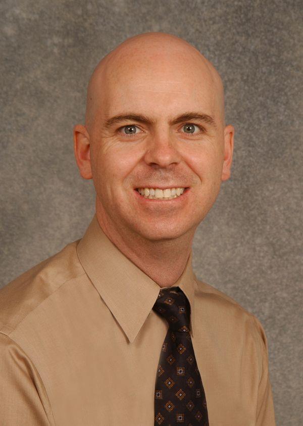
Todd Hankinson, MD
Division Head
Pediatric Neurosurgery
Children's Hospital Colorado
Professor of Neurosurgery and Pediatrics
University of Colorado School of Medicine
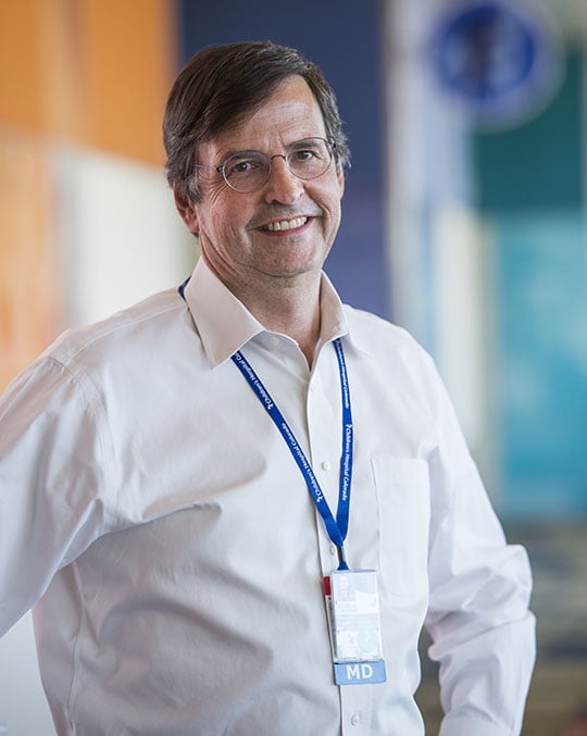
Nick Foreman, MD
Seebaum/Tschetter Chair of Pediatric Neuro-Oncology
Center for Cancer and Blood Disorders
Children's Hospital Colorado
Professor
Pediatrics-Hematology/Oncology and Bone Marrow Transplantation
University of Colorado School of Medicine
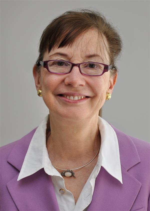
BK Kleinschmidt-DeMasters, MD
Professor
Pathology
University of Colorado School of Medicine
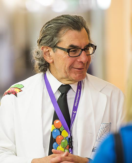
Michael Handler, MD
Associate Surgeon-in-Chief
The Neuroscience Institute
Children's Hospital Colorado
Chairman
Department of Neurosurgery
University of Colorado School of Medicine
Jean Mulcahy Levy, MD
Pediatric neuro-oncologist
Center for Cancer and Blood Disorders
Children's Hospital Colorado
Associate professor
Pediatric Hematology/Oncology and Bone Marrow Transplantation
University of Colorado School of Medicine