How can artificial intelligence models use personalized medicine to improve outcomes for children with craniosynostosis?
An infant’s skull is held together by flexible tissues called sutures, which expand to allow bone growth and accommodate a rapidly growing brain. In children with craniosynostosis, sutures fuse prematurely, causing skull malformation and raising risks of elevated intracranial pressure, vision changes and developmental delays. Treatment for craniosynostosis can have varied results because surgeons may not have data that could help them accurately predict patients’ cranial growth. Now, craniofacial surgery experts at Children’s Hospital Colorado are employing artificial intelligence to create personalized, data-driven models that improve outcomes.
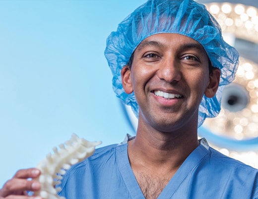
Antonio R. Porras, PhD, pediatric plastic and reconstructive surgery research director at Children’s Colorado and assistant professor of biostatistics and informatics at the University of Colorado School of Public Health, leads this work. Dr. Porras’ team uses artificial intelligence (AI) software to fuel photogrammetry, which takes images of a child’s head from various angles. The resulting anatomical representation not only helps diagnose cranial abnormalities sooner, but also helps predict their progression — all with extraordinary accuracy.
“When we get a patient in the clinic now, we have methods that can tell us exactly what part of the cranium is abnormal, by how much it is abnormal and how much less volume the brain has in a specific area of their head,” Dr. Porras says. “And that is extremely important for surgeons to plan their treatments.”
AI modeling for craniofacial surgery
AI modeling is a novel approach for craniofacial surgeons, but the data sustaining this software is years in the making. A decade ago, Children’s Colorado’s craniofacial clinic co-director Brooke French, MD, launched an image database intended to improve evaluations for patients with craniofacial differences. The database has nearly 1,500 images, including CT images and 3D photograms of craniosynostosis patients before and after surgery. This database was then extended by Dr. Porras’ team to include CT images of patients without craniosynostosis and those with intracranial hypertension, or elevated head pressure — a potential complication of untreated craniosynostosis. Both sexes are represented across the dataset.
“The work of my team was to leverage all the data that Dr. French acquired and build a tool that would inform us toward the best course of action for every patient,” Dr. Porras says.
These images provide a wide range of references for comparison that guide artificial intelligence modeling. This helps quantify even subtle anomalies with greater accuracy. “We can finally start to know what the right surgery is, for the right patient, at the right time, which will help us personalize their medicine really nicely,” Dr. French says.
Improving craniofacial surgery accuracy
Today, this technology transcends the limitations of previous cranial models in multiple ways. For one, the noninvasive approach may eliminate the need for multiple CT scans to evaluate craniofacial differences, which expose children to harmful radiation and often require sedation.
Instead, these new methods can use fewer CT scans to quantify individual details, such as bone shape, thickness and mineral density. Additionally, they use 3D photogrammetry to create models that account for variability in head shape between the sexes. Both methods help improve surgical accuracy.
Additionally, Dr. Porras and his team can use the tool to predict changes during a child’s life up to 10 years of age, which is when the most cranial growth occurs.
These personalized predictions are the key to preventing complications that can be life-changing for craniosynostosis patients. “We can enable our surgeons to take action before there is an actual problem with the child’s development,” Dr. Porras says.
Since these research methods are less focused on a specific pathology and more focused on understanding abnormal patterns of cranial growth at large, this approach to prevention may extend to children with other craniofacial conditions.
Dr. Porras was recently awarded an R01 grant from the National Institutes of Dental and Craniofacial Research, which he will use to build the first quantitative reference for local head volume development in craniosynostosis patients. Specifically, this reference will illuminate the correlations between regions of the head that are underdeveloped, and those experiencing overgrowth — a key metric for planning effective treatment.
This research will also create methods and tools for evaluating, comparing and predicting growth after different treatments, as well as potential relapses, which will help surgeons identify the best course of action.
Collaborating to understand mechanisms of craniofacial disease
The Craniofacial Clinic at Children’s Colorado leads the nation with these AI innovations, but it wouldn’t be possible without such strong multidisciplinary collaboration. Drs. French and Porras’ advancements in care are a result of partnerships with the neurosurgery and craniofacial surgery departments at Children’s Colorado, as well as the biostatistics and informatics, medical genetics and craniofacial biology departments at the University of Colorado. This approach was vital not only in creating the computational methods and software, but also fostering ease of use and implementation for clinical teams.
“It’s really hard finding a team that can do all these things together,” Dr. Porras says. “We’ve been very lucky here that we have been able to create a multidisciplinary team between the university and the hospital.”
The goal of this collaboration is to both advance understanding of the developmental mechanisms of craniofacial disease, and to use this knowledge to create tools that surgeons can easily use for faster, more accurate treatment. Currently, Dr. Porras is asking surgeons to try out the tool and provide feedback to evaluate its usability. That way, surgeons can analyze a patient’s head, understand their exact condition and create a plan for treatment — all in real-time.
Featured Researchers

Antonio R. Porras, PhD
Research Director
Pediatric Plastic and Reconstructive Surgery
Children's Hospital Colorado
Assistant professor
Biostatistics and Informatics, Colorado School of Public Health
University of Colorado School of Medicine
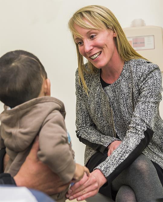
Brooke French, MD, FACS
Co-director, craniofacial surgery
Center for Children's Surgery
Children's Hospital Colorado
Director of the Cosmetic Program
Associate professor of Plastic and Reconstructive Surgery
University of Colorado School of Medicine


 720-777-0123


