Key takeaways
-
The most widely used approaches for evaluating fetal heart rhythms have limits.
-
Our fetal cardiologists were the first to modify the superior vena cava and the ascending aorta (SVC/AAo) technique.
-
The new method, called the InnV/Ao Doppler technique, improves views of the retrograde atrial systolic flow and evaluation of fetal arrhythmia.
Research background: There are limitations in existing methods of assessing fetal arrhythmia
Fetal rhythm abnormalities, which include irregular fetal heart rates, occur in up to 2% of pregnancies and account for 10 to 20% of referrals to fetal cardiologists. Echocardiography is typically used to determine if the fetal heart arrhythmia is benign or if there is a pathological abnormality. During an echocardiogram, fetal heart rhythm is typically assessed using ultrasound tools called M-mode and pulsed spectral Doppler.
Simultaneously recording the pulsed Doppler flow profile in the superior vena cava (SVC) and the ascending aorta (AAo) is more accurate for atrioventricular (AV) and ventriculoatrial (VA) time measurements than M-mode.
Although this pulsed Doppler technique improves accuracy, there are several limiting factors. To be reliable in sampling the low velocity SVC atrial flow and simultaneous AAo, the Doppler signal needs an ideal angle of insonation (approaching 0 degrees) and low wall motion filter (cannot be set too high). Otherwise, the A-wave in the SVC may not be detected.
Research methods: Modifying the way we evaluate arrhythmia in fetus
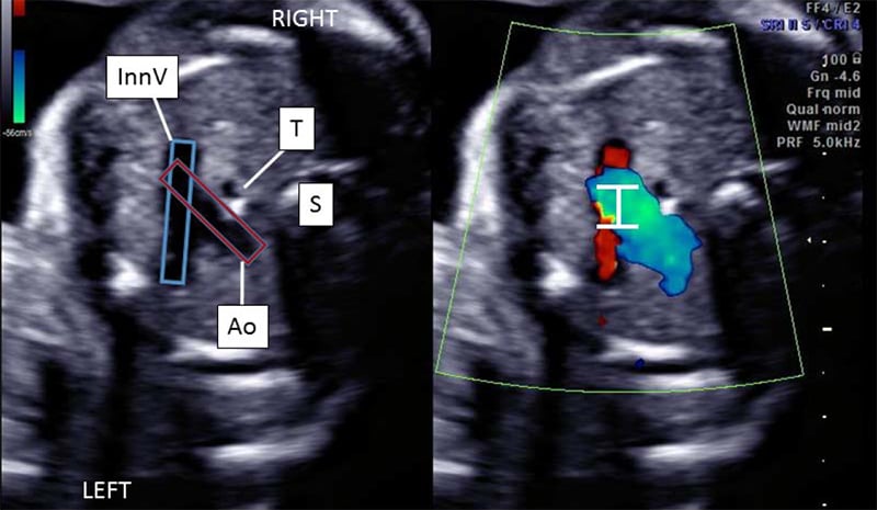
The team in the Fetal Cardiology Program at the Colorado Fetal Care Center at Children's Hospital Colorado, modified the SVC/AAo technique to improve the examination of fetal arrhythmias and evaluating fetal arrhythmias. They simultaneously recorded the pulsed wave Doppler signals in the innominate vein (InnV) and transverse aortic arch (Ao) from an axial view of the fetal thorax.
By employing a four-chamber view in a horizontal position, the InnV is easily identified by sweeping cephalad to the three-vessel trachea view. The InnV is located immediately superior to the transverse arch, and can be positioned in parallel to the ultrasound beam for clear and precise spectral Doppler sampling of the venous A-wave signal.
Research results: An improved view of the retrograde atrial systolic flow
When the Doppler sample volume is widened, blood velocities in both the InnV and the Ao can be simultaneously recorded. (See Figure 1.)


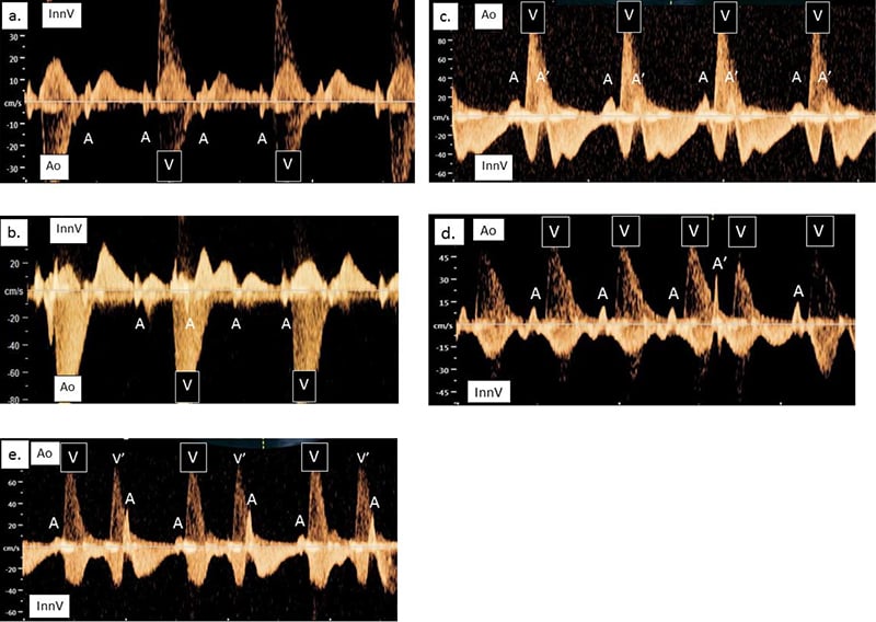
The InnV/Ao method provides a near 0-degree angle of insonation of the InnV to optimize visualization of the retrograde atrial systolic flow. (See Figure 2 for fetal arrhythmias detected with the method.)


Research conclusion: Our InnV/Ao Doppler technique is an acceptable modification of the SVC/AAo technique
Children's Colorado is the first to use simultaneous Doppler recordings of the InnV and Ao from an axial plane to assess fetal cardiac arrhythmias. The InnV/Ao Doppler technique is an accurate method to assess fetal heart rhythm and can be regularly performed in the clinical setting.


 720-777-0123


