720-777-0123


Key takeaways
-
Jane Stremming, MD, Laura Brown, MD, and Paul Rozance, MD, and colleagues at the Perinatal Research Center are studying the IGF-1 growth hormone, which stimulates growth in some fetal organs.
-
The fetus needs more than pro-growth signaling pathway activation to grow; it also needs a supply of the building blocks from which cells are made.
-
Study findings suggest IGF-1 may promote more efficient utilization of nutrients for growth, rather than increasing the nutrients available for growth from the placenta.
Insulin-like growth factor-1 (IGF-1) is an important growth hormone for the fetus. Low circulating fetal IGF-1 is associated with fetal growth restriction (also called intrauterine growth restriction) and high circulation of the hormone promotes fetal overgrowth. Both are linked to pregnancy complications.
Findings from previous studies about IGF’s role in fetal growth
In humans:
- A strong positive correlation with IGF-1 umbilical cord blood concentrations and birth weight in pregnancies
- IGF-1 receptor gene mutations demonstrate intrauterine growth restriction
In mouse models:
- IGF-1 and IGF-1 receptor gene inhibition leads to fetal growth restriction in mouse models.
- Skeletal muscle is the primary organ impacted by IGF-1.
- IGF-1 helps produce myoblasts and turn them into muscle fibers in postnatal muscle cells.
- IGF-1 receptor gene knockout mice have smaller muscles with lower numbers of myocytes.
- Overexpression of IGF-1 in adult mice and infusion of recombinant human IGF-1 into adult rats leads to increased muscle size.
In sheep:
IGF-1 supplementation improves fetal growth in fetal sheep
- Infusion can increase whole body fetal weight.
- Size of select organs, including the heart, are increased.
- Intravenous or intra-amniotic infusions improve fetal growth rates.
Although IGF-1 is known to promote fetal growth and skeletal muscle growth, little is known about its role in regulating placental blood flow, nutrient transfer and nutrient availability to support fetal growth. What is known includes:
- IGF-1 can stimulate uptake of glucose and amino acids in vitro in human placental trophoblast cells.
- The effect of in vivo IGF-1 infusion on placental expression and nutrient transport activity is variable.
- IGF-1 intra amniotic infusion to growth restricted sheep fetuses yield higher placental mRNA expression of amino acid transporters and may have increased nutrient transport to the fetus, supporting growth.
- IGF-1 infusion reduced placental clearance of amino acid and glucose analogs in normally growing, late gestation fetal sheep.
- Infusion of IGF-1 over several hours increased glucose and nitrogen concentrations in the fetus, suggesting increased fetal and placental uptake of glucose and amino acids.
- Recombinant human IGF-1 infusions over several hours led to lower protein breakdown and leucine oxidation rates.
In this study, researchers sought to determine the effect of a one-week fetal infusion of LR3 IGF-1 on net uterine and umbilical nutrient uptake rates, fetal nutrient availability and fetal protein kinetic rates in late gestation fetal sheep.
Research methods: a week-long infusion of LR3 IGF-1 to measure fetal growth
Infusion and metabolic study
The study, led by Dr. Jane Stremming, Dr. Laura Brown and Dr. Paul Rozance and colleagues at the Perinatal Research Center at Children’s Hospital Colorado, included two cohorts. Catheterized fetuses from late gestation pregnant sheep were randomly assigned to receive an intravenous infusion of the synthetic protein long arginine 3 IGF-1 (LR3 IGF-1; n = 8) or saline (SAL; n = 8) for one week.
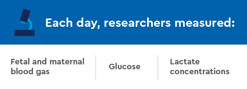


On the final day of infusions, a metabolic tracer study was performed, where uterine and umbilical blood flow, nutrient uptake rates and fetal protein kinetic rates were measured.
In total, eight animals each completed saline or LR3 IGF-1 infusions.
Skeletal muscle analysis
To measure myoblast replication and myofiber hypertrophy, study authors used muscle biopsy samples from a secondary cohort of animals from a similar study of LR3 IGF-1 infusion into late gestation fetal sheep.
Immunohistochemistry
A subset of five SAL and seven LR3 IGF-1 fetuses were used for an immunohistochemical muscle analysis.
Research results: organ growth increased, but not from stimulated nutrient uptake
Fetal and placental weights
There was no statistical difference in fetal weight between the saline group and the LR3 IGF-1 group. In LR3 IGF-1 group, fetal heart, adrenal gland, and spleen weights were higher and insulin concentrations were lower. After normalizing individual organ weights to fetal weight, heart and spleen weights remained significantly higher in LR3 IGF-1, while the normalized brain weight was significantly lower.
Fetal blood gas, substrate and hormone measurements
In the LR3 IGF-1 group, endogenous IGF-1 concentrations, insulin concentrations, oxygen content, oxygen saturation, glutamate and isoleucine concentrations were all lower compared to the saline group.
Uterine and umbilical blood glow, nutrient uptake rates and nutrient/oxygen quotients
Uterine and umbilical blood flow and umbilical uptake rates of glucose, lactate and oxy- gen were similar between the groups. Umbilical amino acid uptake rates were lower in LR3 IGF-1, as were fetal concentrations of multiple amino acids.
Fetal protein metabolism
In LR3 IGF-1 group, net leucine uptake by the fetus and flux of leucine into the fetal blood from the placenta was lower.
Myoblast proliferation and myofiber cross-sectional area
Fetal protein kinetic rates were similar. LR3 IGF-1 skeletal muscle had higher myoblast proliferation.
Key result
Study authors found a one-week infusion of LR3 IGF-1 into late gestation fetal sheep promoted organ-specific growth, but the primary physiological mechanism was not by stimulation of nutrient uptake rates by the uteroplacental unit or the fetus.
Research discussion: how IGF-1 could be used to promote fetal growth
In contrast to their hypothesis, study authors found umbilical uptake rates of glucose and lactate to the fetus were similar to the saline group after one week of LR3 IGF-1 infusion, and the umbilical uptake rate of amino acids was lower in LR3 IGF-1.
Other considerations by study authors
- Findings indicate a decreased placental transport of amino acids to the fetus after a week-long infusion of LR3 IGF-1, but further studies are needed to examine the mechanisms responsible for this difference.
- Lower fetal insulin concentrations likely contributed to lower umbilical transport of amino acids to the fetus, but it also requires further investigation.
- Amino acid/oxygen quotient was lower in LR3 IGF-1; despite normal glucose/oxygen and lactate/oxygen quotients, the total nutrient/oxygen quotient was lower in LR3 IGF-1, which could mean lower oxidative metabolism of amino acids.
- IGF-1 may initiate growth pathways such as MAP kinase (MAPK) or phosphoinositide 3-kinase (PI3K), leading to cellular proliferation.
- Birth weight and umbilical cord concentrations of IGF-1 are associated with each other, but larger fetuses following infusion of IGF-1 are not a consistent finding in animals that are not growth restricted. There may be limits to fetal growth when the animal has a normal growth trajectory, or limits in substrate availability when IGF-1 concentrations are elevated that also may prevent overgrowth.
- It is unknown if fetal body weight and organ growth initially increased but then slowed over time during the course of the IGF-1 infusion. Also, it is not known if umbilical uptake of amino acids was consistently lower throughout the infusion or if it also increased and then decreased over time.
Research conclusion: IGF-1 may promote more efficient utilization of nutrients for growth
Study authors determined that a one-week infusion of LR3 IGF-1 into late gestation fetal sheep lowered umbilical uptake rates of amino acids, fetal arterial amino acid and insulin concentrations and fetal oxygen content. Despite this reduction in nutrient supply to the fetus, they found that LR3 IGF-1 still promoted the growth of several organs and proliferation of myoblasts and maintained fetal protein metabolism.
Authors interpret these findings to mean that LR3 IGF-1-treated fetuses can effectively utilize the available nutrients and oxygen to support organ-specific growth, rather than IGF-1 infusion stimulating placental blood flow or nutrient transfer to the fetus.
Further studies are needed to examine the specific metabolic pathways responsible for decreased placental transport of amino acids, increased organ growth and increased myoblast proliferation by LR3 IGF-1 and to determine how important it is to maintain insulin, amino acid and oxygen concentrations during LR3 IGF-1 infusion to maximally support growth and protein accumulation.
Featured Researchers

Laura Brown, MD
Neonatologist
Neonatal Intensive Care Unit
Children's Hospital Colorado
Professor
Pediatrics-Neonatology
University of Colorado School of Medicine

Paul Rozance, MD
Neonatologist
Children's Hospital Colorado
Professor of neonatology
University of Colorado School of Medicine

Jane Stremming, MD
Neonatologist
Neonatal Intensive Care Unit
Children's Hospital Colorado
Assistant professor
Pediatrics-Neonatology
University of Colorado School of Medicine