720-777-0123


Key takeaways
-
Our researchers have discovered a minimally invasive method to repair open neural tubes using a material they bioengineered.
-
In mouse models, the reverse thermal gel successfully attached to the skin and was found to be safe for use in fetuses.
-
The study demonstrates the potential to use the new approach as an alternative to existing in-utero repairs of spina bifida.
Research background
In the developing fetus, the brain and spinal cord form from a neural tube. Failure of this tube to completely close causes neural tube defects, such as spina bifida. Up to 10% of infants born with an open neural tube defect die because of complications including brainstem dysfunction, shunt malfunctions and infections.
Babies born with open spina bifida can have significant complications including bowel and urinary incontinence, leg weakness or paralysis and sexual and psychological dysfunction.
In utero spina bifida repair of open neural tube defects is possible, but occurs relatively late (22 to 24 weeks of gestation), and preservation of neurological function is minimal. At the time of the repair, the neural tissue has already been damaged by mechanical trauma, chemical irritation and inflammation associated with exposure to amniotic fluid.
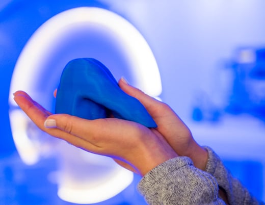
Researchers at the Colorado Fetal Care Center and the Department of Bioengineering at the University of Colorado Denver, Anschutz Medical Campus, are developing an alternative approach. They've created a minimally-invasive repair method using a bioengineered material – a reverse thermal gel – to cover the open neural tube defect at an earlier gestational age.
Reverse thermal gels are biomaterials that change state, from a liquid to a gel (a jello-like substance), with a change in temperature. They are liquid at room temperature and spontaneously assemble into a solid scaffold at human body temperature. Cells or therapeutics can be mixed into a liquid reverse thermal gel, which can then serve as a scaffold for controlled release of therapeutic agents.
Research methods
The researchers chemically synthesized a novel reverse thermal gel, called PSHU-PNIPAAm, and characterized its ultrastructure by scanning electron microscopy, its stability in amniotic fluids and its permeability.
They studied the effects of PSHU-PNIPAAm on basic cellular functions of mouse neonatal fibroblasts, keratinocytes and neurons (the types of cells exposed in open neural tube defects) and they injected the material into mouse embryos in utero.
Research results and discussion
Physical characteristics of PSHU-PNIPAAm:
- Honeycomb or branching structure with variable pore size depending on polymer concentration
- PSHU backbone does not degrade after up to 30 days of exposure to human or sheep amniotic fluid
- Minimal limited and predictable permeability
- Forms a stable gel at temperatures that reflect the intrauterine environment
- Polymer gel is stable in an amniotic fluid environment for two months
PSHU-PNIPAAm is biocompatible with mouse neonatal fibroblasts, keratinocytes and neurons and it supports normal growth of these cells. It is not cytotoxic to these cell types.
Direct microinjection of PSHU-PNIPAAm into an ex utero mouse spina bifida model embryo was technically successful and did not affect the viability of the embryo, which continued to develop in culture. PSHU-PNIPAAm supports cellular migration of mouse embryonic fibroblasts, a proxy measure for wound healing capacity.
Ultrasound-guided injections in utero into the amniotic sac of embryonic mice were successful and compatible with fetal survival and maintenance of pregnancy in 50% of cases. The reverse thermal gel successfully formed a gel and attached to the skin, demonstrating successful in utero suitability as a potential alternative for open neural tube defect closure.
Research conclusions
A novel biomaterial that is liquid at room temperature and changes to a gel at body temperature meets criteria to act as a potential repair material for spina bifida defects. The biomaterial, PSHU-PNIPAAm:
- Possesses limited and predictable permeability
- Is biocompatible with types of cells exposed in neural tube defects
- Is non-toxic
- Has a backbone that is stable in amniotic fluid
- Is compatible with embryos.
In utero application of the biomaterial was technically feasible and did not harm fetuses.
In ongoing studies, the investigators will explore the ability of PSHU-PNIPAAm to act as a scaffold for cellular interaction. They will also make additional chemical modifications to incorporate biomolecules that mimic the extracellular matrix environment of the fetus and enhance cellular activities.
The novel biomaterial PSHU-PNIPAAm will continue to be explored as a repair material for neural tube defects. The potential to use the material in a minimally-invasive approach (direct injection with a fine gauge needle) at earlier gestational ages offers a promising alternative to current in utero repairs for spina bifida.
Funding for this research was provided by the Fetal Health Foundation.