What can 4D MRI reveal about single ventricle flow patterns and how they correlate to clinical outcomes?
The three operations that rebuild the circulatory system for kids with hypoplastic left heart syndrome— the Norwood, the Glenn and the Fontan — have saved thousands of lives. But it’s not a perfect solution. Fontan circulation creates a host of problems that affect every system of the body, but it’s largely a mystery how and why. “This work is a first attempt to push back the veil and understand what happens downstream in these patients after the blood leaves the heart,” says pediatric cardiologist Michael Di Maria, MD. “That’s never really been looked at before using these MRI techniques.”
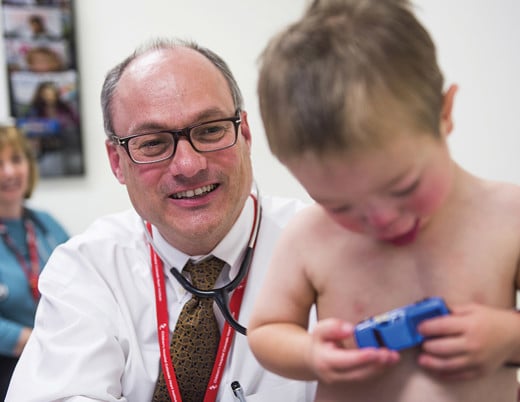
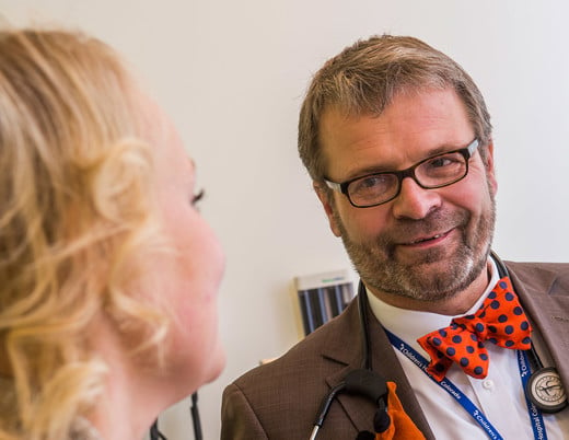
To look at it, Dr. DiMaria partnered with biomedical engineer Michal Schäfer, PhD, who, three years into medical school, boasts a lengthy and growing list of publications for his work in 4D flow MRI, which has produced novel measurements and biomarkers in dozens of diseases affecting the vasculature.
Drs. Schäfer and DiMaria looked at Fontan propagation through two novel lenses: wave intensity analysis and principal component analysis. They then squared their findings against a registry, developed in part by Dr. DiMaria and unique to Children’s Hospital Colorado, that collects a huge amount of prospective data on single ventricle patients during the course of care, allowing them to correlate their measurements with clinical outcomes.
The results lay the groundwork for a new way of understanding Fontan circulation. In fact, a third paper deconstructing some of their earliest findings has the potential to change the way surgeons perform the Norwood.
1. Principal component analysis
The same technique that powers facial recognition software, principal component analysis deciphers morphological patterns based on how often they occur in a data set, such as MRI images.
“Then we correlated those features with functional metrics we already use,” says Dr. Schäfer. “Boom, we got a correlation.”
The flow patterns they identified using MRI associated with the degree of stiffness and dilation in the functional ventricle, which in turn correlated to good or poor patient exercise output, a standard measure of system capacity. Notably, relaxation turned out to be just as important as contraction: A compliant ventricle not only helps to push blood through the system, but to pull blood out of the lungs as it descends back toward the heart.
2. Wave intensity analysis
“The heart and blood vessels have this dance called coupling,” says Dr. Di Maria. “The heart delivers a beat and the blood vessels receive it, and what you want is good cooperation between the two.”
Dr. Schäfer broke that reciprocal action into three basic components: forward compression, backward compression, and forward decompression. Of particular interest was backward compression, referring to the vessels’ ability to receive flow. The stiffer the vessel, the greater the wave. A lead pipe, for example, would have greater backward compression than a rubber hose.
“The patterns that we saw were way different from the control population,” Dr. Di Maria says. “Off the grid.”
Not surprisingly, a lower backward compression wave correlated with better exercise output in the patient data. As a next step, Drs. Schäfer and Di Maria hope to gather real-time exercise data using a specially built supine bike they can roll right up to the MRI.
Another key variable proved to be the stiffness of the aorta, which is surgically reconstructed during the Norwood procedure with a patch. But it wasn’t just the stiffness. It was also the shape.
3. Norwood: brief research report
A close examination of flow through the aorta revealed that hemodynamics were in large part dictated by geometry. Specifically, svelte, tubular aorta performed more efficiently than a wide, cylindrical one — suggesting that a reconstruction mirroring the shape and geometry of the natural aorta is not only a good aesthetic ideal, but is also physiologically worth the effort.
The response was immediate. The Journal of Thoracic and Cardiovascular Surgery published editorials with titles like “Form improves function: the importance of a well-constructed neoaortic arch” and “The perfect arch: A pipe dream that may be achievable!”
“To achieve the ideal Norwood arch reconstruction, we will need to tailor the arch to patient-specific anatomic variations and dimensions,” mused one. “Use of 4-dimensional flow MRI, … may help determine preoperatively the size and shape of an ideal aortic arch reconstruction for a specific patient subtype."
"Ultimately, the importance of any new medical technology is measured by its influence on clinical care,” enthused another. Given the persistent clinical vulnerability of patients with single-ventricle congenital heart disease, 4D MRI has the potential for significant influence on their outcomes.”
Four have been published altogether — so far.
“That kind of response,” Dr. Schäfer says. “It’s kind of unheard of.”
Featured Researchers
Michael Di Maria, MD
Pediatric cardiologist
Children's Hospital Colorado
Associate professor
Pediatrics-Cardiology
University of Colorado School of Medicine
Michal Schafer, PhD
Clinical instructor
Surgery-Cardiothoracic
University of Colorado School of Medicine


 720-777-0123


