720-777-0123


Can doctors use MRI scans and a quantitative risk assessment to alert for serious placenta conditions in pregnant patients?
Currently, doctors rely on ultrasounds to identify potential problems with the placenta in pregnant patients. New studies from Mariana Meyers, MD, Director of Fetal Imaging at the Colorado Fetal Care Center, Children’s Hospital Colorado, and her colleague at the University of Colorado, Erin Englund, PhD, are exploring the benefits of MRI scans and developing a quantitative risk scale to confirm these serious conditions. Their work will not only help expectant patients make more informed decisions about their pregnancy and birth, but it will also better equip care teams when creating treatment plans for those with high-risk placenta complications. This new approach could open doors for more radiologists to feel confident reading these types of images, too, creating better access to this information.
Identifying placenta accreta
For pregnant patients with placenta accreta, where the placenta grows too deep into the uterine wall, a pre-labor diagnosis is essential. This condition inhibits the patient’s ability to have a vaginal delivery and can increase the risk of hemorrhaging. Typically, this condition is caught during a routine pregnancy ultrasound, which triggers the placenta team, a multidisciplinary group of experts across the Anschutz Medical Campus, to come together and formulate the best care plan. Usually, this means scheduling a cesarean section followed by a hysterectomy.
Rather than just relying on ultrasounds to identify conditions like placenta accreta, Drs. Meyers and Englund are advocating for MRIs, so care teams can access more details to implement the best approach to care for the patient.
“Locally, it’s a lot more common to do MRIs, and that is, in large part, because of Dr. Meyers’ expertise,” Dr. Englund says. “She’s advocated for more of these moms to be getting MRIs, so that she can help to define whether they need to have a C-section for their delivery or hysterectomy and go through some of those delivery planning processes.”
Analysis of fetal MRI scans
In their current study, Drs. Meyers and Englund are combining their expertise in hopes of finding a new approach to provide the multidisciplinary placenta team with more information when these conditions are identified and to provide other radiologists the tools to confidently use MRIs in the diagnosis process.
Dr. Meyers’ expertise is extremely specialized — she is one of only two doctors on the Anschutz Medical Campus who reads fetal MRI studies along with David Mirsky, MD, and nearly 300 fetal MRIs were performed at the Colorado Fetal Care Center last year alone. Her expertise is complemented by Dr. Englund, an assistant professor in the University of Colorado’s department of radiology, who uses her expertise in bioengineering to focus on the technical aspects.
“A lot of times, my work is theoretical application,” Dr. Englund explains. “Being able to work with Dr. Meyers on this project helps me see the clinical impact, and seeing how it could impact an individual patient is a really motivating aspect of the research for me.”
And for Dr. Meyers, the feeling is mutual: “Vice versa for me. If I know something is needed clinically, but I don’t know how to achieve it from a technical standpoint, that’s where Dr. Englund chimes in.”
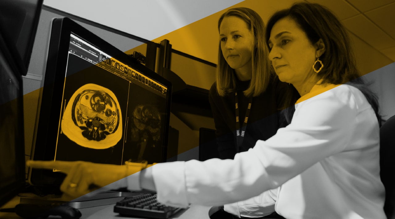
Together, they developed a quantitative risk scale to analyze MRI scans and confidently determine whether a patient meets the threshold for placenta accreta. They then confirm the severity of it to help dictate what should go in their care plan. The team does this by taking MRI scans from pregnant patients and circling the areas on the placenta that appear unusual. Then, Dr. Englund runs her analysis on the identified spots to evaluate the differences of the placental appearance in normal regions and abnormal regions. The pair presented their process and initial pilot study findings to the Radiological Society of North America last November.
Now in the image analysis phase of the study, they are currently having other doctors identify spots to circle on the MRI scans to see if confidence in reading these images and identifying the spots can accurately extend beyond Dr. Meyers’ expertise.
“These studies are very hard to read,” Dr. Meyers says. “Sometimes, when the findings are very subtle, even for people who see these types of studies often, it is still hard. If there is a way we can provide more assurance in those moments, I think that would help because then the radiologist would be clearer in how the report is written.”
Risk assessment in twin-twin transfusion syndrome
The pair is taking the lessons they’ve learned from the placenta accreta study to launch a new study exploring the placenta and risk assessment in twin-twin transfusion syndrome, or TTTS. TTTS is a rare pregnancy disorder where identical twins share the same placenta, which can cause one of the twins to receive more amniotic fluid than the other. If the balance between the twins becomes too uneven, intervention is needed to increase chances of survival.
The Colorado Fetal Care Center, which offers state-of-the-art testing and innovative care for this rare condition, granted seed funding to Drs. Meyers and Englund to apply the same quantitative risk scale concept to provide more detailed information in TTTS cases.
The team is analyzing MRI scans to evaluate not just the placenta structure, but also various functions like blood flow to each twin, oxygen levels and how readings differ between the maternal and fetal aspects. They are scanning the twins’ brains and placentas before and after selective fetoscopic laser photocoagulation (SFLP), the procedure designed to bring balance to both fetuses. This is a procedure the Colorado Fetal Care Center has performed more than 300 times in the past decade with a survival rate for both twins that surpasses the national average. With images before and after this procedure, not only can Drs. Meyers and Englund compare the twins to each other, but they can compare images before and after SFLP to see what they can learn about fetal neuro development. They are developing a score sheet to determine how the brain structure might change.
“I think this really does have the potential to help us understand the pathophysiology and the things that are changing in twin-twin transfusion syndrome, and then to potentially help dictate clinical care in the future,” Dr. Englund says. “As we get the twin-twin transfusion project up and going, I think that’s going to be a really exciting area of research and a productive place for us to contribute — not only to advancing the knowledge, but also being able to potentially help individual patients.”
Featured Researchers

Mariana Meyers, MD
Director of Fetal Imaging
Vice Chief of Operations, Pediatric Radiology
Children’s Hospital Colorado
Associate professor
Pediatric Radiology
University of Colorado School of Medicine