720-777-0123


Key takeaways
-
Controversy surrounds the use of the appendix for diagnosis of TCA.
-
Colorectal researchers categorized the presence of ganglion cells in the appendix to determine its reliability as a histological diagnosis tool for TCA.
-
Use of the appendix for TCA diagnosis yielded 100% sensitivity specificity, positive predictive value and negative predictive value.
-
If no ganglion cells are found in the appendix, even neonates can be reliably diagnosed with TCA – but it should never be used as the sole diagnostic tool.
Background
Hirschsprung disease (HD) is a congenital condition characterized by the lack of ganglion cells in portions of the intestine (aganglionosis). Total colonic aganglionosis (TCA) is a rare and severe form of HD, affecting one in every 50,000 live births.
Rectal biopsies are the traditional diagnostic method for HD, using frozen sections intra-operatively to determine the extent of colonic aganglionosis. If the appendix is found to be aganglionic, a diagnosis of TCA is made.
There have been questions surrounding this practice, including case reports of aganglionic appendix specimens in a ganglionic colon. The limited understanding of normal appendix histology is a cause for criticism; there is a lack of unaltered specimens available for research and frozen biopsies can be unreliable.
Researchers in the International Center for Colorectal and Urogenital Care at Children’s Hospital Colorado sought to address the lack of data on the role of appendix biopsy in the diagnosis of TCA and the lack of understanding of normal appendix histology using a case series.
Methods
This combined retrospective and prospective study was performed at Children’s Colorado, a leading referral center for HD and other pediatric colorectal conditions.
The retrospective component included permanent histological specimens from 2015 to 2020:
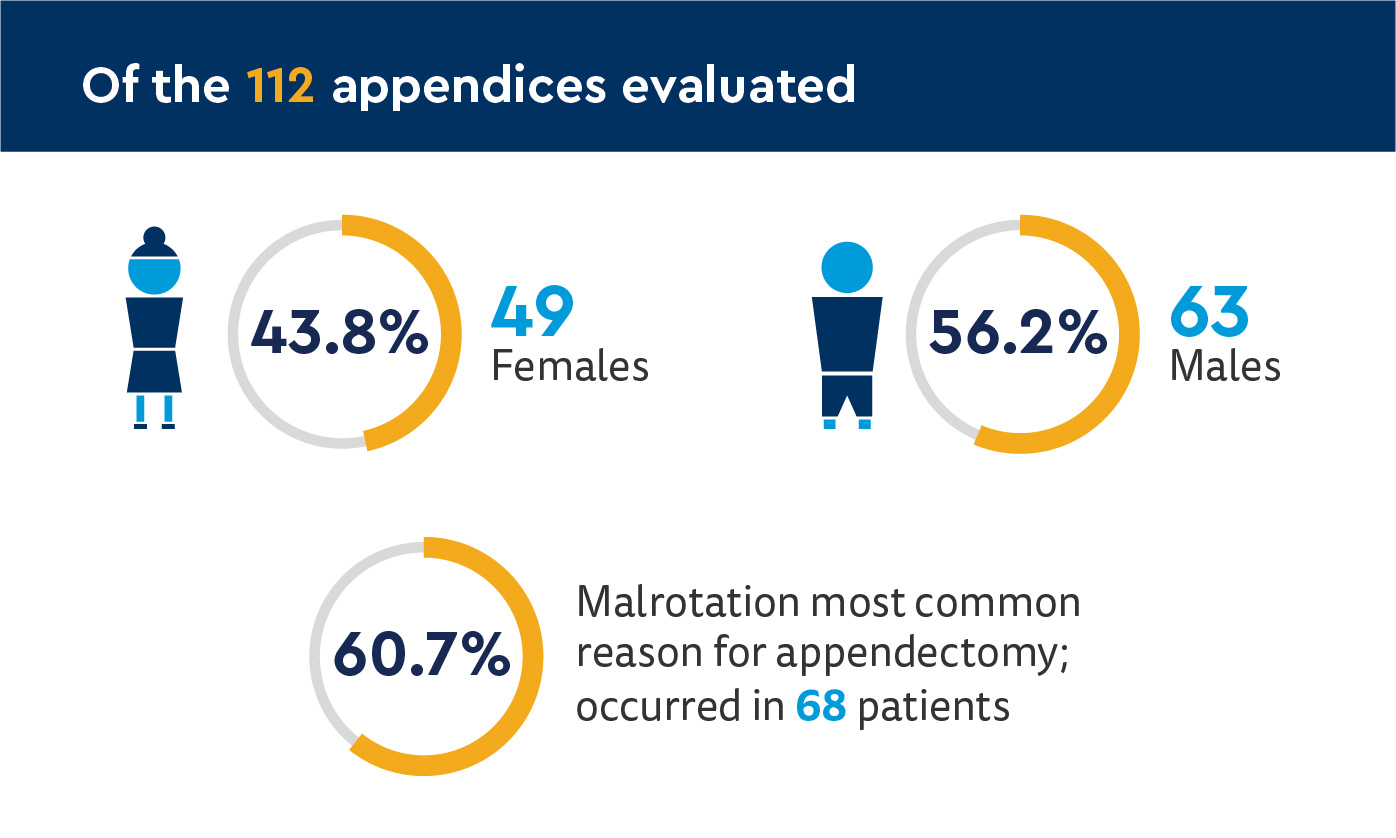
- Normal appendices removed during appendectomy for malrotation
- Falsely presumed appendicitis
- Patients with short-segment HD or TCA
The prospective component included permanent specimens of the appendix from Malone procedures from March 2020 to May 2021.
Study authors also recorded patient demographic information, comorbid conditions, reason for operation and age at appendix specimen collection.
Results: Ganglion cells found in all patients without TCA; none in patients with TCA
HD incidence
There were 14 patients with HD in the study:
- 5 with rectosigmoid HD
- 4 incidental appendectomies during small bowel obstruction surgery
- 1 Malone procedure
- 9 with TCA
- 6 pull-through procedures
- 3 ostomies (awaiting future pull-through)
Pathologic analysis
- 103 (92%) patients had ganglion cells in histologic specimens (none had TCA)
- 9 (8%) patients did not have ganglion calls (all had TCA)
- No difference in presence of ganglion cells by gender
- 28 (25%) patients were less than 30 days old
- 5 (17.8%) with no ganglion cells in appendix specimens
- Groups of ganglion cells found in the submucosa, within the muscular wall of appendices of non-TCA infants
- No ganglion cells, positive fibers in muscularis mucosae lamina propria found in appendices of TCA patients

Discussion and conclusion
Study authors emphasized all TCA patients also had confirmatory rectal biopsies showing lack of ganglion cells, and the appendix should not be used as the sole diagnostic indicator for TCA or used in isolation.
In addition, while the difference was not significant, the average number of ganglion cells in HD patients was less than in control appendices. This underscores the need for judicious histologic examination of the appendix, which is only possible with permanent specimens prior to the final determination of aganglionosis.
They also noted the use for the diagnosis of TCA of a distal appendix specimen measuring approximately 1 cm could preserve the appendix of patients with other diagnoses who may need a Malone procedure or Mitrofanoff procedure.
In conclusion, the absence of ganglion cells in the distal 1 cm of the appendix in a routine histological examination is a reliable tool for diagnosing TCA on permanent section, including in neonates.
Featured Researchers

Michael Arnold, MD
Medical Director of Anatomic Pathology
Pathology and Laboratory Services
Children's Hospital Colorado
Associate professor
Pathology
University of Colorado School of Medicine

Andrea Bischoff, MD
Pediatric surgeon
International Center for Colorectal and Urogenital Care
Children's Hospital Colorado
Professor
Surgery-Peds Surgery
University of Colorado School of Medicine

Luis De La Torre-Mondragon, MD
Pediatric colorectal surgeon
International Center for Colorectal and Urogenital Care
Children's Hospital Colorado
Associate professor
Surgery-Peds Surgery
University of Colorado School of Medicine

Alberto Peña, MD
Founding Director
International Center for Colorectal and Urogenital Care
Children's Hospital Colorado
Professor emeritus
Surgery