Key takeaways
-
It was previously unknown if Schwann cells/glia are a source for intrinsic or extrinsic neurons of the human GI tract.
-
Researchers examined distal rectal tissue from 10 children with normal GI tracts and 48 children with anorectal malformations (ARM) to determine if there are neurons in the human distal GI tract with features of both Schwann cell/glia and neurons.
-
Researchers found dual expression of Schwann cell/glial and neuronal markers in rectal and perirectal neurons in humans, providing the first evidence for a model of Schwann cell-derived neurogenesis in the innervation of the human GI tract.
Research background: examination of distal rectal tissues
Recent studies raised questions about the cell source of enteric neurons in the human GI tract, when research found 20% of these neurons in the mouse colon originated from a Schwann cell precursor. Researchers in the nationally recognized Digestive Health Institute at Children’s Hospital Colorado sought to find evidence of a similar process in humans.
Research methods: analyzing distal rectal tissue
For the control group, researchers studied distal rectal tissue taken at autopsy from 10 children who died of unrelated illnesses and had no gastrointestinal disease or anorectal malformations (ARM).
To study ARM, the researchers analyzed 48 surgical specimens consisting of the most distal colonic/rectal segments removed as part of pull-through surgery to repair the colon and pelvic floor. Of these specimens, six had neurons within the extrinsic rectal innervation. These were further investigated with immunohistochemistry for neuronal and Schwann cell/glial markers.
Of the cadaveric samples, six were female, and ages ranged from a 41-gestational-week fetus to 14 years of age. Of the 48 ARM specimens, 31 were female, ranging from 11 days old to 4 years at time of surgery.

Research results: GLUT1-positive extrinsic innervation in distal colonic/rectal specimens
Researchers found evidence of dual expression of Schwann cell/glial and neuronal markers in the rectal and perirectal neurons in both the control and ARM samples, supporting a model of Schwann cell-derived neurogenesis in the innervation of the human GI tract. Perirectal tissue from control and ARM contained GLUT1-positive extrinsic nerves, many containing neurons.
Additionally, researchers found that the neurons in perirectal nerves were 61% larger in ARM samples; these data suggest that GLUT1-positive extrinsic nerves are distributed in perirectal tissue and the rectum itself in both normal development and different types of imperforate anus malformation but tend to be larger and more plentiful in the latter.
Research results: investigation of Schwann cell markers
To investigate the possible co-expression of Schwann cell/glial markers in perirectal neurons, researchers examined the expression of glial biomarkers SOX10, MPZ, CDH19, and PLP1 in both the control and ARM samples.
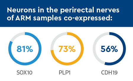
Neurons in the perirectal nerves of ARM samples co-expressed:
- SOX10 (81%)
- PLP1 (73%)
- CDH19 (56%)

Conversely, neuronal co-expression of PLP1 and CDH19 was observed in less than 2% of control samples.
Research conclusion: a model of Schwann cell-derived neurogenesis in the human GI tract
The study authors’ findings suggest cells of a Schwann cell/glial lineage are a source of intrinsic and extrinsic neurons in the human GI tract. They found evidence of dual expression of Schwann cell/glial and neuronal markers in rectal and perirectal neurons in the samples. This is the first evidence supporting a model of Schwann cell-derived neurogenesis in the innervation of the human GI tract, to the knowledge of study authors.
If Schwann cell-derived neurogenesis occurs in human rectal enteric neurodevelopment, it is possible Schwann cell precursors might be induced to form neurons in the aganglionic rectum of patients with Hirschsprung disease as an alternative or adjunct to contemporary surgical management. Hirschsprung disease is a congenital condition that involves missing nerve cells in part, or all of, the large intestine, making it difficult for infants to pass stool.
Future research in this area will improve knowledge of the normal and abnormal development of the enteric nervous system and its response to injury, providing opportunities to develop new therapies for diseases of the GI tract.
Featured Researchers

Michael Arnold, MD
Medical Director of Anatomic Pathology
Pathology and Laboratory Services
Children's Hospital Colorado
Associate professor
Pathology
University of Colorado School of Medicine

Jaime Belkind-Gerson, MD
Pediatric neurogastroenterologist
Children's Hospital Colorado
Associate professor, section head
Pediatrics-Gastroenterology, Hepatology and Nutrition
University of Colorado School of Medicine

Andrea Bischoff, MD
Pediatric surgeon
International Center for Colorectal and Urogenital Care
Children's Hospital Colorado
Professor
Surgery-Peds Surgery
University of Colorado School of Medicine

Mark Lovell, MD
Pediatric pathology specialist
Pathology and Laboratory Services
Children's Hospital Colorado
Professor
Pathology
University of Colorado School of Medicine

Alberto Peña, MD
Founding Director
International Center for Colorectal and Urogenital Care
Children's Hospital Colorado
Professor emeritus
Surgery


 720-777-0123


